

QUANTITATIVE FLUORESCENT POLYMERASE CHAIN
REACTION (QFPCR) IN THE PRENATAL AND POSTNATAL
DIAGNOSIS OF THE MOST FREQUENT ANEUPLOIDIES
Macek M Sr1,*, Krebsova A1,2, Brou_ková M1, Matj_ková M1,
Machatková M1, Diblík J1, Sperling K2, Vorsanova S3, Kutsev S4,
Zerova S5, Arbuzova S6, Chudoba D1, Novotná D1
*Corresponding Author: Associate Professor Milan Macek, Sr., MD, PhD, Centre of Reproductive Genetics, Institute of Biology and Medical Genetics, University Hospital Motol, Charles University, 2nd Medical School, V uvalu 84, Prague, CZ 150 06, Czech Republic; Tel.: +4202-2443-3534; Fax: +4202-2443-3525; E-mail: pavel.roubic@lfmotol.cuni.cz
page: 87
|
|
RESULTS AND DISCUSSION
In Table 4 are documented the first results concerning the parental and meiotic origin of trisomy 21 in the studied German, Russian and Ukrainian families, together with the analysis of the epidemiological data. They have been prepared for the case-control study of trisomy 21 and other most frequent aneuploidies, to examine the impact of the Chernobyl accident, different lifestyles and environmental and genetic factors on meiosis I and II errors, and of their parental origin. The presented data demonstrate that, in the Russian and Ukrainian families, there is no different proportion of paternal non-disjunction estimated for 5-10% [39] relative to the proportion of maternally derived cases. The proportion of the meiosis I errors varies between 63% (Berlin) and 77% (Rostov, Kiev), thus around 70% in agreement with the studies of Petersen and Mikkelsen [40] and Hassold and Sherman [41], except for the families from Moscow (93.9%). Further analysis of more families, of the proportion of errors of meiosis I and II, of maternal and paternal origin together with the results of the case-control and epidemiological studies, are under way in order to find out the possible impact of the Chernobyl accident, and other factors that might endanger the meiosis process.
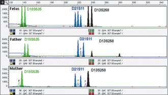
Figure 1. The 1:1:1 height (area) peak ratio of the D21S11 marker demonstrates the trisomy 21 in the affected fetus. Comparison of the length of the PCR fragments in father and mother revealed a maternal origin of the extra chromosome 21. The retained heterozygosity of D21S11 in the fetus indicates that the non-disjunction occurred in meiosis I.
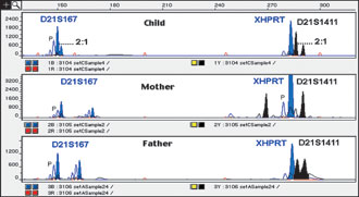
Figure 2. The 2:1 height (area) peak ratio of the D21S167 and D21S1411 markers demonstrates the trisomy 21 in the affected child. Comparison of the length of the PCR fragments in father and mother revealed a paternal origin of the extra chromosome 21. The heterozygosity of D21S21411 and D21S167 in father is reduced to homozygosity in the affected child, indicating, that the non-disjunction occurred in meiosis II.
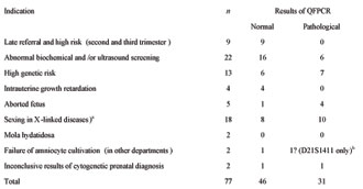
Table 2. Possible indications for quantitative fluorescent polymerase chain reaction (QFPCR) examination.
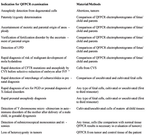
Table 3. Results of prenatal QFPCR examination of aneuploidies of chromosomes 13, 18, 21, X and Y (2000-2003). (All QFPCR results were confirmed by cytogenetic analysis except the cases with mola hydatidosa and abortions because of cell/tissue degeneration and thus cultivation.)
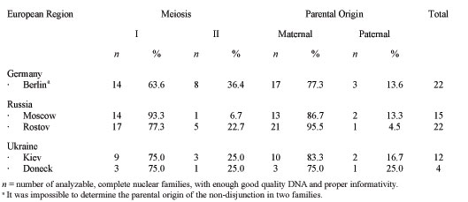
Table 4. Parental and meiotic origin of trisomy 21 in German, Russian and Ukranian families, ascertained by quantitative fluorescent polymerase chain reaction (QFPCR).
|
|
|
|

 |
Number 28
Vol 28 2025 Supplement |
Number 27
VOL. 27 (2), 2024 |
Number 27
VOL. 27 (1), 2024 |
Number 26
Number 26 VOL. 26(2), 2023 All in one |
Number 26
VOL. 26(2), 2023 |
Number 26
VOL. 26, 2023 Supplement |
Number 26
VOL. 26(1), 2023 |
Number 25
VOL. 25(2), 2022 |
Number 25
VOL. 25 (1), 2022 |
Number 24
VOL. 24(2), 2021 |
Number 24
VOL. 24(1), 2021 |
Number 23
VOL. 23(2), 2020 |
Number 22
VOL. 22(2), 2019 |
Number 22
VOL. 22(1), 2019 |
Number 22
VOL. 22, 2019 Supplement |
Number 21
VOL. 21(2), 2018 |
Number 21
VOL. 21 (1), 2018 |
Number 21
VOL. 21, 2018 Supplement |
Number 20
VOL. 20 (2), 2017 |
Number 20
VOL. 20 (1), 2017 |
Number 19
VOL. 19 (2), 2016 |
Number 19
VOL. 19 (1), 2016 |
Number 18
VOL. 18 (2), 2015 |
Number 18
VOL. 18 (1), 2015 |
Number 17
VOL. 17 (2), 2014 |
Number 17
VOL. 17 (1), 2014 |
Number 16
VOL. 16 (2), 2013 |
Number 16
VOL. 16 (1), 2013 |
Number 15
VOL. 15 (2), 2012 |
Number 15
VOL. 15, 2012 Supplement |
Number 15
Vol. 15 (1), 2012 |
Number 14
14 - Vol. 14 (2), 2011 |
Number 14
The 9th Balkan Congress of Medical Genetics |
Number 14
14 - Vol. 14 (1), 2011 |
Number 13
Vol. 13 (2), 2010 |
Number 13
Vol.13 (1), 2010 |
Number 12
Vol.12 (2), 2009 |
Number 12
Vol.12 (1), 2009 |
Number 11
Vol.11 (2),2008 |
Number 11
Vol.11 (1),2008 |
Number 10
Vol.10 (2), 2007 |
Number 10
10 (1),2007 |
Number 9
1&2, 2006 |
Number 9
3&4, 2006 |
Number 8
1&2, 2005 |
Number 8
3&4, 2004 |
Number 7
1&2, 2004 |
Number 6
3&4, 2003 |
Number 6
1&2, 2003 |
Number 5
3&4, 2002 |
Number 5
1&2, 2002 |
Number 4
Vol.3 (4), 2000 |
Number 4
Vol.2 (4), 1999 |
Number 4
Vol.1 (4), 1998 |
Number 4
3&4, 2001 |
Number 4
1&2, 2001 |
Number 3
Vol.3 (3), 2000 |
Number 3
Vol.2 (3), 1999 |
Number 3
Vol.1 (3), 1998 |
Number 2
Vol.3(2), 2000 |
Number 2
Vol.1 (2), 1998 |
Number 2
Vol.2 (2), 1999 |
Number 1
Vol.3 (1), 2000 |
Number 1
Vol.2 (1), 1999 |
Number 1
Vol.1 (1), 1998 |
|
|
|