

FAMILIAL UNBALANCED REARRANGEMENTS
OF CHROMOSOME 5 DUE TO A MATERNAL
BALANCED TRANSLOCATION 5;9
Sukarova-Angelovska E, Kocova M*
*Corresponding Author: Professor Dr. Mirjana Kocova, Pediatric Clinic, Medical Faculty, Vodnjanska 17, 1000 Skopje, Republic of Macedonia; Tel: +389-111-713; Fax: 389-129-027; E-mail: ozonunit@unet.com.mk
page: 73
|
|
CLINICAL REPORT
The proband was a 20-day-old male newborn of unrelated parents, both aged 40; he was the seventh child in the family. The first three children (one girl and two boys) died at different ages (3 months, 1 year and 3 years, respectively). No data on the cause of death in these children were available. According to the parents, however, all showed developmental delay, and one physically resembled the proband. The cause of the developmental delay was never determined.
The pregnancy with the proband was normal and the baby was born in the 38th gestational week. He was small for the gestational age (1800gr/46cm). After delivery he had to be resuscitated because of fetal distress. After stabilization in the intensive care unit, elucidation of the facial dysmorphism was requested. The baby had a characteristic weak cry, microcephaly, broad forehead, thin and sparse hair, hypertelorism, antimongoloid-slanted small eyes, and lateral placement of inner canthi with short palpebral fissures. Facial dysmorphism also included broad nose, small mouth, thin lips, small tongue, and micrognathia. The ears were low-set and poorly developed. The baby had a short neck, slender body, narrow shoulders and chest, and club feet. The fingers of the hands were relatively long, simian crease was present bilaterally. The nails were hypoplastic on the fingers, and especially on toes (Fig. 1A).
Routine laboratory investigations were normal, except anemia (Hb 10.1 g/dL) and slight hypoproteinemia (total proteins 58 gr/L) due to poor feeding. Ultrasonographic examination of the heart, performed due to systolic murmur, showed a ventricular septal defect into the membranaceous part of the septum. Ultrasonographic examination of the brain raised a suspicion of absence of corpus callosum. Ultrasonographic examination of the kidneys was normal. The baby had swallowing difficulties and nutrition was performed by nasal catheter. Esophagography did not show any structural defect of the esophagus, however, dysfunction of the swallowing act was confirmed. Episodes of milk aspiration were frequent, and the baby suffered several aspiration pneumonias.
The mother of the child was a 40-year-old woman with mild dysmorphic features: flat forehead, protruding eyes, divergent strabismus. Her mental status was within the normal range. She had had surgery because of thyroid carcinoma 3 years prior to this pregnancy, and has been on thyroxin therapy ever since. Her facial appearance could be attributed to the thyroid disease.
The proband's sister was a 14-year-old girl who had previously been diagnosed with cerebral palsy. She had a profound developmental delay, being unable to sit and walk. She could hardly speak or communicate, even with close members of her family. Truncal hypotonia was present with significant stiffness of the great joints. She had had repeated seizures since infancy, despite the anti-convulsive therapy. Her facial dysmorphism included microcephaly, hypertelorism, long and up-slanting palperal fissures, long philtrum, and pointed mandible. Her limbs were slender, with arachnodactylia. She had kyphoscoliosis caused by long lasting immobility. No visceral abnormalities were recorded in this girl (Fig. 1B). Two other girls in the family, aged 10 and 7 years, respectively, were normal, without dysmorphic features or developmental delay. The family pedigree is presented in Fig. 2.
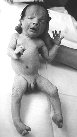

Figure 1. A) The proband with deletion 5p. B)The probands sister with trisomy 5p
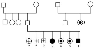
Figure 2. Family pedigree (1*: the proband with CdCS; 2*: the proband's sister with trisomy 5p; 3*, 4* and 5*: mother and two sisters, carriers of the translocation).
|
|
|
|

 |
Number 27
VOL. 27 (2), 2024 |
Number 27
VOL. 27 (1), 2024 |
Number 26
Number 26 VOL. 26(2), 2023 All in one |
Number 26
VOL. 26(2), 2023 |
Number 26
VOL. 26, 2023 Supplement |
Number 26
VOL. 26(1), 2023 |
Number 25
VOL. 25(2), 2022 |
Number 25
VOL. 25 (1), 2022 |
Number 24
VOL. 24(2), 2021 |
Number 24
VOL. 24(1), 2021 |
Number 23
VOL. 23(2), 2020 |
Number 22
VOL. 22(2), 2019 |
Number 22
VOL. 22(1), 2019 |
Number 22
VOL. 22, 2019 Supplement |
Number 21
VOL. 21(2), 2018 |
Number 21
VOL. 21 (1), 2018 |
Number 21
VOL. 21, 2018 Supplement |
Number 20
VOL. 20 (2), 2017 |
Number 20
VOL. 20 (1), 2017 |
Number 19
VOL. 19 (2), 2016 |
Number 19
VOL. 19 (1), 2016 |
Number 18
VOL. 18 (2), 2015 |
Number 18
VOL. 18 (1), 2015 |
Number 17
VOL. 17 (2), 2014 |
Number 17
VOL. 17 (1), 2014 |
Number 16
VOL. 16 (2), 2013 |
Number 16
VOL. 16 (1), 2013 |
Number 15
VOL. 15 (2), 2012 |
Number 15
VOL. 15, 2012 Supplement |
Number 15
Vol. 15 (1), 2012 |
Number 14
14 - Vol. 14 (2), 2011 |
Number 14
The 9th Balkan Congress of Medical Genetics |
Number 14
14 - Vol. 14 (1), 2011 |
Number 13
Vol. 13 (2), 2010 |
Number 13
Vol.13 (1), 2010 |
Number 12
Vol.12 (2), 2009 |
Number 12
Vol.12 (1), 2009 |
Number 11
Vol.11 (2),2008 |
Number 11
Vol.11 (1),2008 |
Number 10
Vol.10 (2), 2007 |
Number 10
10 (1),2007 |
Number 9
1&2, 2006 |
Number 9
3&4, 2006 |
Number 8
1&2, 2005 |
Number 8
3&4, 2004 |
Number 7
1&2, 2004 |
Number 6
3&4, 2003 |
Number 6
1&2, 2003 |
Number 5
3&4, 2002 |
Number 5
1&2, 2002 |
Number 4
Vol.3 (4), 2000 |
Number 4
Vol.2 (4), 1999 |
Number 4
Vol.1 (4), 1998 |
Number 4
3&4, 2001 |
Number 4
1&2, 2001 |
Number 3
Vol.3 (3), 2000 |
Number 3
Vol.2 (3), 1999 |
Number 3
Vol.1 (3), 1998 |
Number 2
Vol.3(2), 2000 |
Number 2
Vol.1 (2), 1998 |
Number 2
Vol.2 (2), 1999 |
Number 1
Vol.3 (1), 2000 |
Number 1
Vol.2 (1), 1999 |
Number 1
Vol.1 (1), 1998 |
|
|