GORLINS SYNDROME: CASE REPORT AND MANAGEMENT PROTOCOL
Rosti RO1,*, Aktas I2, Kayserili H1, Yalcın S2
*Corresponding Author: Dr. Rasim O. Rosti, Department of Medical Genetics, Istanbul Medical Faculty, Istanbul University, 34390 Capa, Istanbul, Turkey; Tel./Fax: +90-212-534-84-40; E-mail: ozgurrosti@yahoo. com
page: 61
|
|
PROPOSED MANAGEMENT PROTOCOL
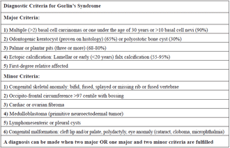
The diagnostic criteria for GS and frequencies of the major findings are reported in Table 1 [5].The presence of multiple basal cell nevi, odontogenic keratocysts, palmar/ plantar cysts and a characteristic facial appearance fulfill the criteria for the diagnosis in our patient.
The follow-up of patients with GS is important because proper patient care leads to early diagnosis and treatment of neoplasms of multiple tissues, thereby improving the quality of life of patients. We here propose a management protocol as outlined in Table 2.
Skin findings should be monitored closely at 6-monthly intervals, especially after puberty, since the rate of malignant transformation increases with full-blown puberty [1]. Since sunlight is a promoter of such malignancies, sun screen precautions should be strongly recommended [6].
Odontogenic keratocysts may appear as early as the fifth year of life [7]. We recommend that annual dental screening should start at the age of 8 years. If there is a jaw cyst then the frequency of dental visits should be adjusted accordingly.
Table 1. Diagnostic criteria list and the frequencies of major findings (5)
FOLLOW-UP OF GORLINS SYNDROME Table 2.
Surveillance of neoplasms, other than skin neoplasms, is crucial for the survival of the patient. Medulloblastoma typically presents during the first 2 years of life as opposed to 7 to 8 years in the general population, and the likelihood of developing a medulloblastoma is unlikely after 7 years of age [5]. Due to the fact that craniospinal irradiation results in hundreds of basal cell carcinomas of the irradiated field, routine scanning with computed tomography or excessive use of radiography is not recommended [8]. There is no evidence that magnetic resonance imaging is of value. Clinical neurological examination at 6-month intervals is the choice of surveillance until 3 years of age, when the clinical evaluation could be rescheduled to once a year. In older children, headaches, vomiting, dizziness, or a change in personality or habits reported by the parents, could be pivotal hints for detection of central nervous system neoplasms.
The musculoskeletal manifestations consisting of bifid, fused or splayed ribs; missings ribs or vertebrae; lack of segmentation of cervical or upper thoracic vertebrae, ectopic calcification of various sites and pseudocystic lytic bone lesions, should only be documented if the diagnosis is uncertain, since radiation exposure promotes the occurrence of skin malignancies [5,8].
Cardiac fibromas and ovarian fibromas occur at a higher frequency in GS than expected [5]. Since cardiac fibromas, likely to cause clinical problems, may be present from very early in raphy is optional unless there are suggestive symptoms. The same approach applies to the possibility of chylous or lymphatic cysts of the mesentry [1].
The ocular problems encountered in GS include congenital cataract(s), microphthalmia, orbital cysts, coloboma of the iris, choroid and optic nerve, stra-bismus and nystagmus [3]. An ophthalmologic examination at the time of diagnosis is recommended. Regular ophthalmologic examinations are recommended if there is an abnormality that necessitates follow-up. In summary, we highly recommend the advised follow-up protocol of GS for better management of complications, and for early detection of tumors that may necessitate early intervention.
|
|
|
|

 |
Number 28
Vol 28 2025 Supplement |
Number 27
VOL. 27 (2), 2024 |
Number 27
VOL. 27 (1), 2024 |
Number 26
Number 26 VOL. 26(2), 2023 All in one |
Number 26
VOL. 26(2), 2023 |
Number 26
VOL. 26, 2023 Supplement |
Number 26
VOL. 26(1), 2023 |
Number 25
VOL. 25(2), 2022 |
Number 25
VOL. 25 (1), 2022 |
Number 24
VOL. 24(2), 2021 |
Number 24
VOL. 24(1), 2021 |
Number 23
VOL. 23(2), 2020 |
Number 22
VOL. 22(2), 2019 |
Number 22
VOL. 22(1), 2019 |
Number 22
VOL. 22, 2019 Supplement |
Number 21
VOL. 21(2), 2018 |
Number 21
VOL. 21 (1), 2018 |
Number 21
VOL. 21, 2018 Supplement |
Number 20
VOL. 20 (2), 2017 |
Number 20
VOL. 20 (1), 2017 |
Number 19
VOL. 19 (2), 2016 |
Number 19
VOL. 19 (1), 2016 |
Number 18
VOL. 18 (2), 2015 |
Number 18
VOL. 18 (1), 2015 |
Number 17
VOL. 17 (2), 2014 |
Number 17
VOL. 17 (1), 2014 |
Number 16
VOL. 16 (2), 2013 |
Number 16
VOL. 16 (1), 2013 |
Number 15
VOL. 15 (2), 2012 |
Number 15
VOL. 15, 2012 Supplement |
Number 15
Vol. 15 (1), 2012 |
Number 14
14 - Vol. 14 (2), 2011 |
Number 14
The 9th Balkan Congress of Medical Genetics |
Number 14
14 - Vol. 14 (1), 2011 |
Number 13
Vol. 13 (2), 2010 |
Number 13
Vol.13 (1), 2010 |
Number 12
Vol.12 (2), 2009 |
Number 12
Vol.12 (1), 2009 |
Number 11
Vol.11 (2),2008 |
Number 11
Vol.11 (1),2008 |
Number 10
Vol.10 (2), 2007 |
Number 10
10 (1),2007 |
Number 9
1&2, 2006 |
Number 9
3&4, 2006 |
Number 8
1&2, 2005 |
Number 8
3&4, 2004 |
Number 7
1&2, 2004 |
Number 6
3&4, 2003 |
Number 6
1&2, 2003 |
Number 5
3&4, 2002 |
Number 5
1&2, 2002 |
Number 4
Vol.3 (4), 2000 |
Number 4
Vol.2 (4), 1999 |
Number 4
Vol.1 (4), 1998 |
Number 4
3&4, 2001 |
Number 4
1&2, 2001 |
Number 3
Vol.3 (3), 2000 |
Number 3
Vol.2 (3), 1999 |
Number 3
Vol.1 (3), 1998 |
Number 2
Vol.3(2), 2000 |
Number 2
Vol.1 (2), 1998 |
Number 2
Vol.2 (2), 1999 |
Number 1
Vol.3 (1), 2000 |
Number 1
Vol.2 (1), 1999 |
Number 1
Vol.1 (1), 1998 |
|
|
|